Thoracocentèse et pose d’un drain thoracique chez les bovins

Auteurs
Résumé
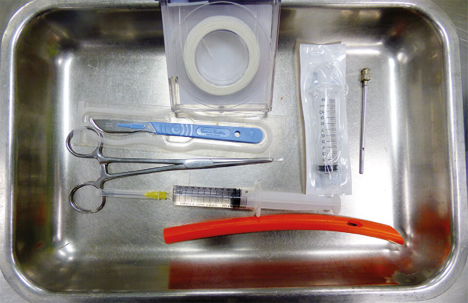
La pose d’un drain thoracique sur les bovins est un acte chirurgical relativement simple à réaliser et qui peut permettre d’obtenir une bonne amélioration clinique. L’intervention est réalisée sur animal debout (au besoin légèrement tranquillisé). La peau et les muscles intercostaux sont d’abord incisés à la lame de bistouri. Puis, le drain thoracique est introduit dans la cavité thoracique grâce à un mandrin métallique à bout pointu permettant de perforer la plèvre pariétale. Le drain est alors raccordé à une valve d’Heimlich ou à un prolongateur de perfusion et une seringue. Les muscles et la peau sont ensuite suturés et le drain est fixé à la peau grâce à la technique du lacet chinois. La pose du drain peut être précédée d’une échographie qui permettra de choisir au mieux le site de ponction, et d’une thoracocentèse afin de caractériser au mieux la nature de l’anomalie (pneumothorax, pyothorax, hémothorax traumatique, épanchement suite à une tumeur affectant les plèvres ou les poumons, …). Ainsi, les résultats de la thoracocentèse, au chevet de l’animal (couleur, odeur, abondance du liquide obtenu, ou présence de gaz), ou consécutifs aux examens de laboratoire (cytologie, bactériologie), permettent de choisir le type de drainage souhaité pour la cavité pleurale. Les principales complications possibles sont une hémorragie (rare), une hypotension (parfois fatale) ou infection.
Abstract
Placing a thoracic drain in cattle is a relatively simple surgical procedure that can provide good clinical improvement. The operation is performed on standing animals (if necessary, slightly sedated). The skin and intercostal muscles are first cut with a scalpel blade. Then, the thoracic drain is introduced into the thoracic cavity using a metal mandrel with a pointed end allowing the parietal pleura to be perforated. The drain is then connected to a Heimlich valve or to an infusion extension line and a syringe. The muscles and skin are then sutured and the drain is fixed to the skin using the Chinese lace technique. An ultrasound can be performed to allow the best choice for the puncture site and the position of the drain. A thoracocentesis can help identify the type of anomaly (pneumothorax, pyothorax, traumatic haemothorax, effusion following a tumour affecting the pleura or the lungs, etc.). The results of an immediate thoracocentesis (colour, odour, abundance of the liquid obtained, or presence of gas), or after laboratory tests (cytology, bacteriology), make it possible to choose the best type of drain for the pleural cavity. The main complications are haemorrhage (rare), hypotension (sometimes fatal) or infection.


